Brain Tumor Treatment using Ion Beams
Slide Show

Heidelberg Ion Beam Therapy Center
The Heidelberg Ion Beam Therapy Center HIT at Heidelberg University Hospital is a unique facility for the treatment of tumors worldwide due to its innovative technology.

Irradiation facilities & accelerator at HIT
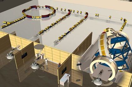
The irradiation facilities and accelerator at the Heidelberg Ion Beam Therapy Center HIT are hidden many meters under the surface and behind thick walls and also covered by a hill of earth that is seven meters high.

Directors at HIT
Prof. Dr. Dr. Jürgen Debus, Medical Director (l.), and Prof. Dr. Thomas Haberer, Head of Research and Technology, work closely at the Heidelberg Ion Beam Center HIT.

Beam guide and treatment tables
This graphic shows the beam guide and the treatment tables at the Heidelberg Ion Beam Therapy Center HIT. HIT has two horizontal treatment tables and a gantry treatment table.

Horizontal treatment table
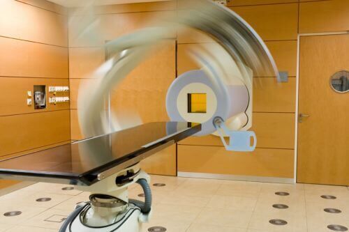
Horizontal treatment table with robot-controlled patient gurney and rotating computed tomography scanners.

Treatment Table in Gantry
Treatment table in the gantry, where the beam can hit the patient at every angle due to the movable gantry.

The Gantry
The gantry is a huge, pivoting beam guide, with the aid of which the patient can be irradiated from all sides. This unique structure worldwide is made of steel, weights 670 tons, is 25 meters long, has a diameter of 13 meters and is three floors high.

Explantation of Procedure
The procedure is thoroughly explained to the patient prior to treatment at the Heidelberg Ion Beam Therapy Center HIT

Individual Irradiation Plan
An individual irradiation plan is drawn up for every patient: This photo shows the dosage distribution overlaid over the CT scan.

© Siemens
Positioning of Patient
Patient prior to treatment with an ion beam. He is positioned precisely with the aid of a laser beam. So that the movements of the body do not influence the treatment results during the irradiation, a synthetic mask is prepared for every patient to hold his head in place. Photo: Siemens

Monitoring during irradiation
X-ray control room for the gantry treatment place. The patient is x-rayed once again, before the irradiation begins. During the treatment, a physician and a medical-technical radiology assistant monitor the irradiation of the patient, which can last up to 30 minutes, depending on the medical condition.

Millimeter-Precise Tailored Beams
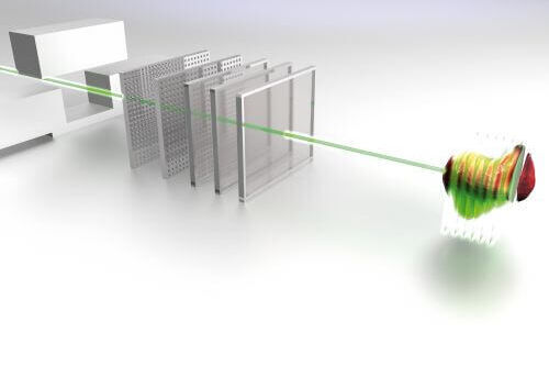
At HIT, a never before reached precision in the 3D irradiation of tumors is now possible due to a special irradiation method, the so-called "intensity-modulated grid-scan procedure". Millimeter-precise tailored beams cover the tumor - like a glove fits the hand - and irradiates the whole shape of the tumor. Photos: University Hospital Heidelberg

